
Throughout Research Week, images submitted by this year’s winners of the School of Medicine Art in Science Competition will be on display in the first floor lobby of Bondurant Hall.
This is an in-person event.
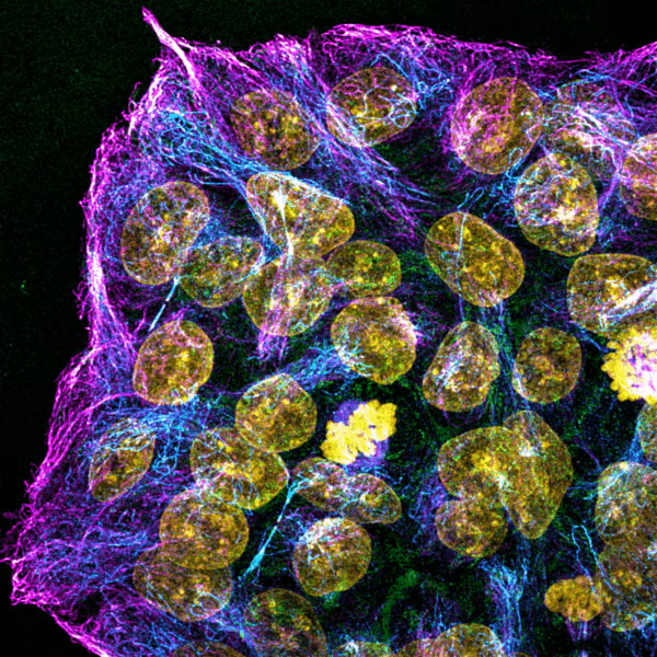
Display image description: “Cracking the Secret Stem Cell Highways Behind Alzheimer’s Disease” was submitted by Chih-Hsuan Hsu and Dr. Sarah Cohen from the Department of Cell Biology and Physiology. One of the winners of the 2025 Art in Science competition. This confocal image reveals the intricate microtubule network within human induced pluripotent stem cells carrying the APOE4 variant, a major genetic risk factor for late-onset Alzheimer’s disease. Immunostaining highlights the “tubulin code” with magenta, cyan, green, and yellow markers, offering a glimpse into how genetic variants shape cellular architecture and function.
2026 Winners
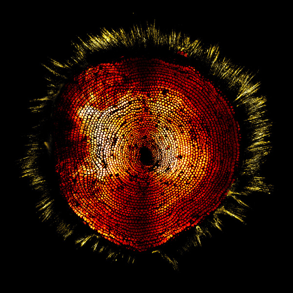
“Foraminifera Sunrise”
Gabriel Kreider & Dr. Klaus Hahn
Department of Pharmacology
A large benthic foraminifera (Amphisorus kudakajimensis) stained with CaMero, biosensor for calmodulin activity
that consists entirely of a small molecule, so it can be used in genetically intractable organisms. Red shows
chlorophyll fluorescence from symbiotic algae (zooxantela) in the calcified shell of the foraminifera. Yellow shows
calmodulin activation in reticulapodia protruding from the shell and attaching to the coverslip. CaMero can
simply be added to extracellular medium or injected in animals to visualize signaling activity, expanding the range
of accessible organisms, and we hope enabling application in humans. Image was acquired with 4x magnification
on a spinning disk confocal microscope.
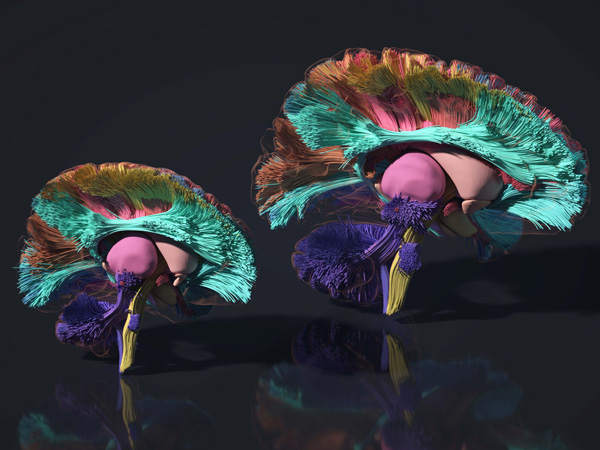
“Mapping the Developing Brain’s Information Highways”
Khoi Huynh, Junjie Zhao, Sahar Ahmad, & Dr. Pew-Thian Yap
Department of Radiology
Using advanced MRI, the Yap Lab visualizes the brain’s information highways as they develop during early life.
The image displays these neural pathways from birth (left) to age five (right), revealing how the brain’s
communication network evolves over time. Mapping this emerging network offers new insight into how the
developing brain builds the foundation for learning, movement, and lifelong health. The data was acquired at
BRIC as part of the UNC/UMN Baby Connectome Project.
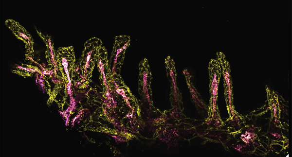
“Two roads diverged in a … villus”
Dr. Hannah Hrncir & Dr. Kathleen Caron
Department of Cell Biology and Physiology
Mouse small intestine (ileum) has been trimmed into thin strips to allow 3D imaging of whole villi using the
Andor Dragonfly Spinning Disk microscope. Blood vasculature is shown in yellow, and lymphatic vasculature is
shown in magenta. Whether a molecule is absorbed through the yellow or magenta routes determines its
journey through the body. For instance, small water-soluble molecules enter the blood circulation (yellow) and
are transported directly to the liver, whereas many larger and lipid soluble molecules are absorbed through
lymphatic circulation (magenta) initially bypassing the liver and entering systemic blood flow. Understanding
how molecules, particularly drugs, are absorbed is therefore critical for predicting where they travel and act in
the body. Image taken within the Microscopy Services Laboratory, part of Carolina Microscopy.